2016年5月14日 星期六

壓瘡定性評估方法。NPUAP 2016年版壓傷分期

傷口大師 游朝慶 醫師

  在2016年4月8-9日的一場超過400位專家出席的共識會上(NPUAP 2016 Staging Consensus Conference,如圖1) (注:NPUAP,美國國家壓瘡諮詢委員會,National Pressure Ulcer Advisory Panel ),大家對於壓瘡的分期及定義這議題交互討論,以及對於爭議部分予以表決,在這會議中,與會者都使用照片來確認每一個新的術語( terminology)。最後NPUAP在4月13日向世界公布這新的壓傷(pressure injury)語言以及其修正後的分期。同時作為美國健保給付依據的OASIS (Outcome and Assessment Information Set,居家照護評估表)也將同步做修正。

clip_image002

圖1. NPUAP 2016 Staging Consensus Conference

  壓傷的分期是用來表示組織受傷的程度。之前臨床工作者嘗試去診斷及確認壓傷的期別所會遇到的問題都在這次被NPUAP修正。

"Pressure injuries are staged to indicate the extent of tissue damage. The stages were revised based on questions received by NPUAP from clinicians attempting to diagnose and identify the stage of pressure injuries. 

NPUAP Staging Task Force News Release 2016.4.13

   NPUAP並且免費提供13個圖示(圖2-1 - 2-13)來表示各期別(http://www.npuap.org/resources/educational-and-clinical-resources/pressure-injury-staging-illustrations/).


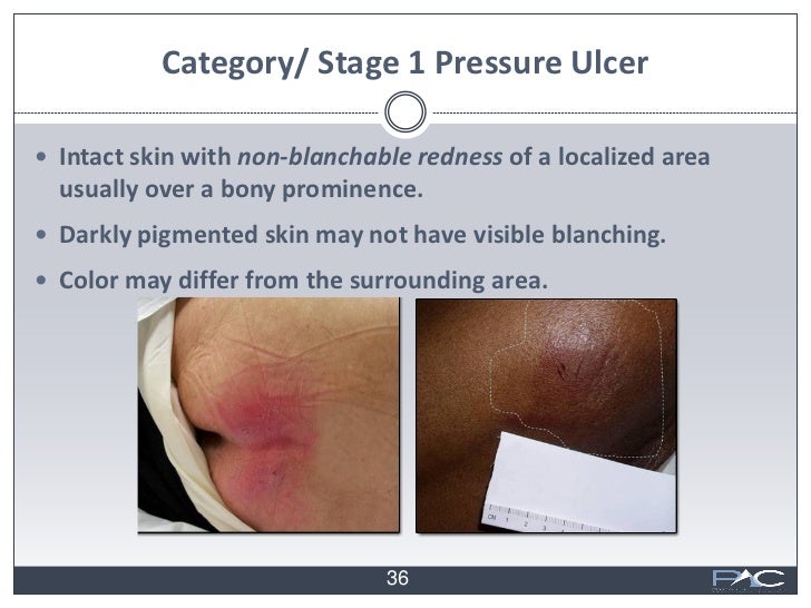
第1期壓傷: 完整的皮膚上有紅斑,給予指壓時不會消失

Stage 1 Pressure Injury: Non-blanchable erythema of intact skin

   在完整的皮膚上看到局部有指壓時不會消失的紅斑(如圖2-4, 圖2-6),但這在皮膚較黑的患者上可能會看不到變化(如圖2-5、圖3),雖然如此,可消失的紅斑、感覺異常、體溫異常及皮膚變得較硬都可能會緊接著出現不會消失的紅斑。然而皮膚顏色的變化並不包括紫色或栗色(maroon)(補充:也就是如瘀青般的顏色),其代表著深層組織傷害(deep tissue injury)。
Intact skin with a localized area of non-blanchable erythema, which may appear differently in darkly pigmented skin. Presence of blanchable erythema or changes in sensation, temperature, or firmness may precede visual changes. Color changes do not include purple or maroon discoloration; these may indicate deep tissue pressure injury.

clip_image004

圖2-1. Healthy skin – Caucasian(健康的皮膚–白種人,高加索人)

clip_image006

圖2-2.Healthy skin – Non-Caucasian(健康的皮膚–有色人種)

clip_image008

圖2-3.Stage 1 Pressure Injury – Caucasian(第1期壓傷–白種人,高加索人)

clip_image010

圖2-4.Stage 1 Pressure Injury – Edema(第1期壓傷–水腫),補充:這圖和上一張圖(圖2-3),我看不出差異性,水腫不是壓傷的症狀,但若患者因營養不良導致水腫、或者因失禁導致皮膚潮濕,則比較容易造成第1至第2期壓傷。

clip_image012

圖2-5.Stage 1 Pressure Injury - Non-Caucasian(第1期壓傷––有色人種)

clip_image014

圖2-6.Blanchable vs Non-Blanchable,給予指壓時會消失的紅斑vs不會消失的紅斑

clip_image016

圖3.如右圖,皮膚較黑的人可能無法判斷有無blanchable,圖片取自David Rokes, Debra Kurtz, WoundRounds: Clinical Reimbursement and Wound Care webinar slides, Health & Medicine, Economy & Finance, Business, Nov 11, 2011 http://image.slidesharecdn.com/woundroundsclinicalreimbursementandwoundcarewebinarslides-111111125701-phpapp02/95/woundrounds-clinical-reimbursement-and-wound-care-webinar-slides-36-728.jpg?cb=1321016386

2016年5月6日 星期五

『壓瘡』已被重新命名為『壓傷』

游朝慶 醫師

   The term “pressure injury” replaces “pressure ulcer” in the NPUAP Pressure Injury Staging System

   就在上個月,4/13/2016,美國國家壓瘡諮詢委員會(NPUAP)宣布將常用的『壓瘡pressure ulcer』這個字改名為『壓傷pressure injury』,因為有些壓瘡如第一度壓瘡及深層組織傷害deep tissue injury其皮膚是完整的。

   此外在這個會議中也決議使用阿拉伯數字來命名第1-4期,而不再使用羅馬數字的第I-IV期。並且也把『疑深層組織傷害』的『疑』字拿掉,表示我們可以有信心地正確診斷為『深層組織傷害』。最後這會議也正式承認兩種因醫療器材導致的傷害,也為壓傷的一種,雖然其不符合「在骨頭突出處」這舊定義,第一種傷害為Medical Device Related Pressure Injury:醫療器材導致的壓傷,如Bi-PAP的氧氣罩導致嘴巴鼻子周圍產生壓傷,這類傷口可以使用NPUAP的分期系統來分期,第二種傷害為Mucosal Membrane Pressure Injury:黏膜壓傷,如氣管內管導致口腔的潰瘍,鼻胃管導致的傷口,其無法再被分期。

   詳見 http://www.npuap.org/resources/educational-and-clinical-resources/npuap-pressure-injury-stages/

而且2016年的『壓傷』也被NPUAP重新定義為

    皮膚或皮膚下面的軟組織受損,其通常位於身體骨頭突出處或與某些醫療器材有關。這些傷害看起來可能皮膚是完整的,或有傷口的或者也許會疼痛。導致傷害的原因為強烈的或持續的受壓、或伴隨著剪力,而這些或許也和微氣候、營養、血循、合併症、及軟組織的狀況有關。

A pressure injury is localized damage to the skin and/or underlying soft tissue usually over a bony prominence or related to a medical or other device. The injury can present as intact skin or an open ulcer and may be painful. The injury occurs as a result of intense and/or prolonged pressure or pressure in combination with shear. The tolerance of soft tissue for pressure and shear may also be affected by microclimate, nutrition, perfusion, co-morbidities and condition of the soft tissue.

    相對於壓瘡2007年NPUAP定義(較簡單)

壓瘡是身體骨頭突出處,因為受到重壓,剪力或摩擦,而導致的皮膚或皮膚下面的脂肪或肌肉組織受損,有很多因素會為導致壓瘡,但這些因素的重要性仍然不明。


參考資料來源:

National Pressure Ulcer Advisory Panel (NPUAP) announces a change in terminology from pressure ulcer to pressure injury and updates the stages of pressure injury http://www.npuap.org/national-pressure-ulcer-advisory-panel-npuap-announces-a-change-in-terminology-from-pressure-ulcer-to-pressure-injury-and-updates-the-stages-of-pressure-injury/

2016年3月20日 星期日

糖尿病患者以骨內氣體及皮下氣腫表現的沙門氏桿菌骨髓炎

  這是今年外科醫學會上發表的第二篇海報,這個案原本應該是骨科的,但他們堅持先處理壞死性筋膜炎,不過也學到皮下有air,不一定就是壞死性筋膜炎,若術前沒有CT,還真診斷不出來。

 今天也終於將blogger上照片顯示不出來的問題解決了,雖然是以很笨的方式重新一張一張貼,但若還是有遺漏的、或需訂正的,麻煩請告訴我,謝謝大家。

DSC03448


糖尿病患者以骨內氣體及皮下氣腫表現的沙門氏桿菌骨髓炎

游朝慶

台南市立醫院 外科


前言:

   當骨頭內在影像學上發現有空氣時(intraosseous gas),但又沒有骨頭傷害時(如骨折或開刀),須強烈懷疑是產氣性骨髓炎(emphysematous osteomyelitis),此外骨髓炎以皮下氣腫(subcutaneous emphysema)來表現的個案,文獻上非常少。下面介紹一個少見的案例,其為一54歲糖尿病患者診斷為左脛骨遠端產氣性骨髓炎,卻以左小腿及大腿的皮下氣腫來表現,組織培養顯示沙門氏桿菌(Salmonella group C2)。

個案報告﹕

   患者54歲男性,原住民,有糖尿病、高血脂、痛風及C肝等病史,於2個月前去南投打獵,因為動物的內臟不好保存,原住民習慣將捕獲的獵物現場剖肚,取出內臟生吃,但隔天就因為左髖部及膝蓋疼痛至本院急診求診,當時發燒到39.4度C、心跳103bpm,WBC正常,CRP:1.93,當時初步診斷為痛風性關節炎,因血糖高達549mg/dL,故因糖尿病控制不良住院。但關節痛仍然未改善,持續發燒,augmentin 無效,改為Ciprofloxacin ,HbA1C高達15.7%,故安排影像學檢查,X光無異常(如圖1):

clip_image002clip_image004

圖1:lt knee X-ray: Soft tissue swelling, anterior knee.

電腦斷層(CT)則可看到左大腿內側有一包空氣,左脛骨遠端骨頭內有一些空氣(圖2)。

clip_image006clip_image008

clip_image010clip_image012clip_image014clip_image016

圖2. CT: fasciitis with cellulitis of left thigh and arthritic arthropathy of bil knees.

  之後患者症狀漸漸改善,而隨後的blood culture培養出兩套 Salmonella group C2,患者於住院一周後出院,診斷為L't knee arthritis, gout arthritis & septic arthritis,Salmonella bacteremia

之後過了兩個月,患者又因左膝疼痛3天,在外面診所以痛風治療無效,到我們急診求診,來的時候,呈現出toxic signs,有點喘、血壓不高、微燒(37.3C),白血球18290並有band form:17%,其他data: CRP: 1.7, Blood gas: pH:7.416, HCO3:14.4, PCO2:22.9, BE:-8.2. Creat:1.7, BUN:49.6, ALT:92, AST:34, CPK:19, uric acid:8.3, sugar:229.

理學檢查為大腿下1/3疼痛,從左側鼠蹊至腳踝都有捻髮音(crepitus),X-Ray顯示左下肢有大量的氣體,懷疑有abscess(圖3),CT顯示左脛骨下1/3有骨髓內氣體,證實有骨髓炎,從左髖部到小腿有大量的皮下氣體,懷疑有壞死性筋膜炎(圖4),因懷疑敗血,建議膝上截肢(AK),但患者拒絕。

clip_image018clip_image020

圖3.Soft tissue swelling with emphysema over the right thigh, knee and lower leg

clip_image022clip_image024clip_image026clip_image028clip_image030clip_image032clip_image034

圖4. CT: intramedullary gas in the distal third of the femur bone, with extensive subcutaneous emphysema

  手術從大腿遠端外側進入,骨頭外側為正常、無膿瘍的表現,我們將脛骨遠端以氣鑽鑽開幾個洞,引流出白色無臭味的膿及氣體,並在左小腿及大腿內側做筋膜切開術,但卻無壞死性筋膜炎的表現。術中的培養結果仍然是Salmonella group C2,但血液培養沒長菌。

clip_image036clip_image038

圖5.OP finding ,第一及第二次手術

  抗生素醫感染科醫師建議使用Antibiotics treatment with Ciprofloxacin 200mg/vial 2 vial Q12H IV + Amoxicillin 1000mg+Clavulanate 200mg 1 vial q8h IVD

在經過兩次手術將傷口縫合後,患者於34天後出院。

結果:

  患者在門診追蹤1個月,並持續口服抗生素治療,傷口狀況良好。

討論:

   患者是台灣的原住民,常與族人一起打獵,並習慣和著鹽巴生吃獵物的內臟,其應該是沙門氏桿菌菌血症的原因。沙門氏桿菌很少會造成骨髓炎。在1981年,Ram等人發表說電腦斷層(CT)發現骨髓內氣體是骨髓炎的徵兆,在顱骨、脊椎、胸骨、肋骨外的骨頭上發現有骨內氣體,則幾乎可以診斷為產氣性骨髓炎,糖尿病及惡性腫瘤為常見的好發因子。因為這種病死亡率相當高(32%)以及常有併發症,尤其是患者同時有糖尿病,故積極的抗生素治療及手術介入是一定要的。


Salmonella Osteomyelitis with Intraosseous Gas and Subcutaneous Emphysema in the Patient with Diabetes

Chao-Chin Yu

Department of Surgery, Tainan Municipal Hospital

Introduction:

The presence of intraosseous gas in the absence of direct communication between bone and air, such as a compound fracture or recent surgery, is highly suggestive of emphysematous osteomyelitis. However there is rare osteomyelitis report with subcutaneous emphysema. We report a case of a 54-year-old male with diabetes mellitus who presented with emphysematous osteomyelitis of the left distal femur and subcutaneous emphysema of left thigh and calf. Specimen cultures in this case revealed a pure growth of Salmonella group C2.

Case Presentation:

The 54-year-old man with past medical history of poor controlled type 2 DM, hyperlipidemia, gout and hepatitis C was admitted through our emergency department due to left knee and hip joint pain for 8 hours. He was tachycardic (103bpm) and febrile (39.4C). Lab data showed normal white blood count, CRP:1.93, but high level of blood glucose level 549mg/dL, and HbA1c:15.7. Musculoskeletal soft tissue computed tomography (CT) showed fasciitis with cellulitis of left thigh and arthritic arthropathy of bil knees. He was diagnosed with gouty arthritis and septic arthritis and was treated with colchicines 0.5mg 1# qd and antibiotics. He was discharge 1week later after the symptoms improve. The following blood culture showed Salmonella group C2. The patient presented again to the emergency department 2months later with ongoing left knee pain for 3days, in vain with treatment of gout by clinic physician. He presented with toxic signs with dyspnea, dropped blood pressure, mild fever(37.3C) and leukocytosis(18290) with band form:17%. Lab data revealed: CRP: 1.7, Blood gas: pH:7.416, HCO3:14.4, PCO2:22.9, BE:-8.2. Creat:1.7, BUN:49.6, ALT:92, AST:34, CPK:19, uric acid:8.3, sugar:229. Physical exam showed local tenderness over lower third of left thigh and extensive crepitus extending from left groin to the left ankle. X-ray showed soft tissue tissue swelling with emphysema over lt thigh, knee and lower leg. Subsequent CT confirmed evidence of osteomyelitis with intramedullary gas in the distal third of the femur bone, with extensive subcutaneous emphysema involving the left hip, thigh and lower leg. Above knee amputation was suggested at first, but was refused by patient.

Through a lateral approach to the distal femur, penetration of bone was initially performed to drain the non-foul whitish intraosseous abscess and gas. The cortex of bone showed intact. Irrigation with H2O2 and debridement were performed. The fasciotomy of left thigh and calf was also done but no necrotizing fasciitis or abscess was found. Intraoperative cultures revealed Salmonella group C2. Blood cultures taken preoperatively showed no bacterial growth.

Antibiotics treatment with Ciprofloxacin 200mg/vial 2 vial Q12H IV + Amoxicillin 1000mg+Clavulanate 200mg 1 vial q8h IVD was suggested by Infection doctor. The wounds were open cared with normal saline moist gauze packing bid. After 2 subsequent surgeries, he was eventually discharged 34 days after admission.

Results:

The patient had been regularly followed up and received oral antibiotics in my OPD for 1months after discharge. The wound condition was good.

Discussion:

This patient is a Taiwanese aborigines. He hunt the boar with his clansman. Then they usually ate the raw organs of boar with salt which may be the source of salmonella bacteremia. Osteomyelitis is a rarely reported manifestation of Salmonella infection. In 1981, Ram et al. were first to describe the radiological finding of intraosseous gas as a sign of osteomyelitis on computed tomography (CT). When seen in the extra-axial skeleton, intraosseous gas is virtually pathognomonic for emphysematous osteomyelitis. Diabetes mellitus and malignancy are common predisposing factors. Aggressive antimicrobial and surgical intervention is required as emphysematous osteomyelitis is associated with high mortality (32%) and significant morbidity, especially in diabetic patients.

2016年3月19日 星期六

併有心臟衰竭的靜脈鬱滯性潰瘍患者

  今天又是全台灣大外科一年一次的大拜拜,順便也去貼了兩張海報。都只是個案報告。

順便測試google blogger部落格的圖片是否可恢復正常顯示(真的快被搞瘋了)

第一篇的中英文介紹如下:

DSC03449DSC03440


併有心臟衰竭的靜脈鬱滯性潰瘍患者
Venous Stasis Ulcers in the Patient with Heart Failure
游朝慶

台南市立醫院 外科


前言:

   靜脈性潰瘍(Venous ulcer),又被稱為鬱滯性潰瘍(stasis ulcer),是最常見的下肢潰瘍原因。其診斷需根據臨床表現,如靜脈不全、皮膚的變化或下肢水腫,其預後不好,常數年不會癒合。我們介紹一個個案,其兩下肢多處潰瘍、疼痛及水腫已四個月未癒合,我們在兩星期內就把潰瘍治療到接近癒合。


個案報告﹕

   62歲退休男性,有慢性心臟衰竭及中風已四年的病史,兩腳多處潰瘍及腫痛已四個月,此次因為兩腳紅腫熱痛,懷疑蜂窩性組織炎(cellulitis)及深層靜脈栓塞(Deep vein thrombosis)而從門診收住院。理學檢查:兩小腿下1/3有色素沉著(hyperpigmentation),嚴重凹陷性水腫(pitting edema),兩小腿紅腫痛,兩足部均可摸到脈搏,在右小腿有一3*4公分的傷口,並有濕性痂皮(wet eschar),在左小腿則有一巨大的15*10公分不規則淺傷口(如圖1),並有大量滲液。胸部有呼吸喘鳴聲(wheezing),實驗室檢查:WBC: 9810, Seg:87, Hb:13.0, plate:313K, BUN/Cr:23.5/1.2, Alb:2.9, Glu:114, CRP:9.25, Alb:2.9, T4:5.07, TSH:4.0756. EKG顯示有First degree AV block. 胸部X-ray: 心臟肥大、肺水腫(pulmonary edema)並有肋膜積水(如圖2)。周邊血管超音波排除了深層靜脈栓塞,但心臟超音波: chamber dilatation and generalized hypokinesis with impair global performance with ejection fraction : 35%.顯示嚴重心臟衰竭。

imageimage

圖1. hyperpigmentation over lower third of both calf, severe pitting edema, swelling, erythema, tenderness over bilateral calf, with chronic ulcer

image

圖2. cardiomegaly, pulmonary edema with pleural effusion

   因此診斷為心臟衰竭導致的下肢靜脈性潰瘍並鬱滯性皮膚炎,根據心臟科醫師建議,便開始使用大量利尿劑及低劑量dopamine療法,傷口則局部使用優碘粉(iodosorb powder),兩小腿抬高並綁上彈性繃帶。治療一禮拜後,wheezing改善後才去開刀房清創傷口(如圖3),患者在住了12天後出院。

imageimage

圖3.手術後,左小腿傷口已有局部癒合


結果

   患者在出院後一禮拜回診,此時傷口已縮小了7成(如圖4)。

最後診斷為CHF with pul edema with Venous stasis ulcers with cellulitis, bil lower legs, C6EsApPr

imageimage

圖4. OPD回診,兩小腿傷口已縮小


討論:

   靜脈不全導致的靜脈性潰瘍常常容易復發,且常常會持續數禮拜到數年,壓迫治療(使用彈性繃帶或彈性襪)是這種疾病的標準治療,治療6個月成功率約3-6成,治療1年成功率約7-8.5成,但傷口治療師永遠要記得引起靜脈潰瘍的其他原因,如肝硬化、心衰竭、深層靜脈栓塞、肥胖、腳外傷後(如骨折或手術)的後遺症、換過人工髖關節或膝關節、服用抗白血病的藥或其他會導致下肢水腫的藥。以這案例而言,利尿劑對於靜脈不全的水腫無效,但若是肝硬化或心衰竭所導致的下肢水腫,則利尿劑效果非常好。


Venous Stasis Ulcers in the Patient with Heart Failure

Chao-Chin Yu

Department of Surgery, Tainan Municipal Hospital

Introduction:

Venous ulcer, also known as stasis ulcer, is the most common etiology of lower extremity ulceration. The diagnosis of stasis ulcer was based on clinical criteria: venous insufficiency, cutaneous signs and/or severe leg edema. This prognosis of venous ulcer is poor. We report a case of bilateral leg chronic ulcers with pain and edema for 4months who nearly heals his ulcer in only 2 weeks.

Case Presentation:

The 62 year-old retired male with past history of chronic heart failure and old CVA for 4years is well until 2014/12/4 when he suffered from progressive swelling, erythema, pain, tire and heat over his bilateral legs. He was admitted via OPD under the initial suspicion of cellulitis and deep vein thrombosis. Physical examination shows hyperpigmentation over lower third of both calf, severe pitting edema, swelling, erythema, tenderness over bilateral calf and palpable pulses in both feet. A ulcer about 3*4cm with wet eschar 100% over right lower leg. The other huge shallow ulcer about 15*10cm with irregular shape and heavy discharge over left calf. Wheezing in chest was also noted. Lab data shows: WBC: 9810, Seg:87, Hb:13.0, plate:313K, BUN/Cr:23.5/1.2, Alb:2.9, Glu:114, CRP:9.25, T4:5.07, TSH:4.0756. EKG: First degree AV block. Chest X-ray: cardiomegaly, pulmonary edema with pleural effusion. Pheripheral vein sono on 12/6 shows no deep vein thrombosis. Cardiac echo on 12/8 shows four chamber dilatation and generalized hypokinesis with impair global performance with ejection fraction : 35%. Then we start lasix with low dose dopamine therapy by the suggestion of CV doctor. Besides, the wounds were treated with iodosorb powder topical use, legs elevation and compression therapy with elastic bandage under the diagnosis of venous stasis ulcers. He also received an operation for debridement on 12/11 after the wheezing improved. He was discharge on 8/16.

Results:

The patient was followed up in my OPD on 12/23. At that time, the huge ulcer of left leg showed about 70% reduction in wound area and near healed.

Discussion:

Venous ulcers are often recurrent, and open ulcers can persist from weeks to many years. Compression therapy is the standard of care for venous ulcers and chronic venous insufficiency. Success rates range from 30 to 60 percent at 24 weeks, and 70 to 85 percent after one year. However, wound care specialists should always keep in mind other possible causes of venous ulcers include liver cirrhosis, heart failure, deep venous thrombosis, obesity, after-effects of leg injury, homolateral artificial hip and knee joints, and consumption of anti-leukaemia or leg-edema-eliciting drugs.

2015年11月2日 星期一

Aquacel-Ag+打臉文(ConvaTec不要看)

游朝慶 醫師
   今天,正研究著一張資料圖表,ConvaTec公司給的,介紹他們家新出的一種敷料aquacel-Ag+ Extra,封面(如圖1)看起來像是發表於傷口雜誌的研究論文,ConvaTec最近在積極推廣宣傳,相信很多人有拿到這一本,若沒有,網路上也有pdf(http://www.convatec.sk/media/files/next-generation-antimicrobial-dressings-aquacel-ag-extra.pdf)。

圖1.封面 (圖片來源: http://www.convatec.sk/media/files/next-generation-antimicrobial-dressings-aquacel-ag-extra.pdf)
   有一種新出來的抗菌敷料不用靠清創,就可以在一天之內將綠膿桿菌的生物膜的活菌總菌落數viable biofilm bacteria(CFU)減少6個零(從30億減到3千,也就是百萬分之一),而在第四天,讓生物膜數量歸零(圖2),這個資料幾乎就是細菌的『明日之後』了,對於有抗藥性的MRSA生物膜也是以每2天讓細菌減少到原本千分之一的速度,在第5天,讓生物膜數量歸零(圖3)。如此的神奇,已經比手術清創的效果還要神了,我每次在開刀房開完清創手術後,頂多只敢宣稱清完9成,需觀察個幾天,若傷口沒改善還是有可能需要再次清創。此敷料一出,大概像我這類做傷口的外科醫師就不用混了,無論如何,總還是覺得怪怪的,因為圖表的細菌單位用的是生物膜內存活細菌的『總菌落數CFU』。

圖2.綠膿桿菌生物膜被Aquacel-Ag+ extra大屠殺,資料來源:P7,
(圖片來源: http://www.woundsinternational.com/media/other-resources/_/1069/files/content_11301.pdf
http://www.convatec.sk/media/files/next-generation-antimicrobial-dressings-aquacel-ag-extra.pdf)


圖3.MRSA葡萄球菌生物膜被Aquacel-Ag+ extra大屠殺,資料來源:P7,
 (圖片來源:http://www.convatec.sk/media/files/next-generation-antimicrobial-dressings-aquacel-ag-extra.pdf)
 

而什麼是CFU呢?

   菌落形成單位/毫升 (colony forming unit/ml;簡稱 cfu/ml),是計算細菌數量的一種方法,其值越高表示樣品中所含的細菌越多。菌落形成單位的計量方式與一般的計數方式不同,一般直接在顯微鏡下計算細菌數量會將活與死的細菌全部算入,但是CFU只計算活的細菌。當單一的活細菌被移植至agar培養皿,經分裂後,其會形成一個菌落。故每一個活細菌,即是由一個菌落形成單位 (Colony forming unit;簡稱 cfu) 所代表。總生菌數是指食品在經過處理,並於一定條件下進行培養後,所得1 g或1 ml檢測樣本中所含的活細菌總數。傳統的方式是將檢體經過連續稀釋(以十的倍數),然後在培養皿培養出可計算菌落數目,接著計算Plate Count。但是這個方法只能計算水中懸浮的微生物planktonic bacteria,或是遷移中的生物膜碎片,大部分的生物膜是會緊密附著於管壁,且尚未成熟的生物膜系統是不會大量移轉到水中的,且每個生物膜碎片常只能培養出單一菌落,最後,據說在傳統的agar培養皿其實培養不出生物膜,因此傳統的總菌落數CFU(應該是cfu/ml)其實不是研究生物膜的好單位。不過也許研究者用了一種特殊的方法,溶解了生物膜的EPS外衣,卻不會傷害膜內的細嫩細菌,把生物膜內的所有細菌釋放出來,再用CFU的方法研究也不一定。就讓我進一步研究其研究方法。
   根據內文,作者發展出一種實驗室的抗菌評估模式(此作者David Parsons還是Aquacel-Ag及Aquacel-Ag+專利的發明家WO2012136968 A1),用來研究Aquacel-Ag+ extra對於傷口中生物膜內細菌的抗菌效果1,2(謎之音:請參考ConvaTec的內部不公開文件:1,2,這句話怪怪的),生物膜在一種低營養的培養基質中,藉著持續攪動48小時,在棉質紗布中生養。接著這生物膜的存在、成熟狀態及形狀被前文所述的雷射掃描共軛焦顯微鏡(Laser Scanning Confocal Microscopy)證實(如圖4)(謎之音:看到這裡似乎都還合理)。接著將含有生物膜的紗布物質轉移到陷在皮革表面的agar培養皿,以模擬傷口的環境(謎之音:漏餡了,agar並不是生物膜的培養環境,),接著將各種敷料(aquacel-Ag及aquacel-Ag+ extra)置於生物膜表面,加水保濕,並以第二層敷料覆蓋(看起來是防水的aquacel foam dressing),在這5天的培養中,每隔一段時間(24小時)就評估敷料對生物膜的抗菌效果(謎之音:到這個步驟為止,作者並沒有以任何方法,如超音波,將剩下殘存的生物膜打碎,以培養生物膜內的細菌,否則一個生物膜就只能培養出一個菌落),接著為了評估生物膜在被破壞後一天便會重建的條件,又緊接著將新鮮的細菌接種在敷料下面的紗布基質上,接著在其後的幾天培養並評估細菌的存活狀況(圖2及圖3)。(謎之音:圖2 MRSA的aquacel-Ag這組沒有歸零,且原本已被aquacel-Ag抑制成拉鋸狀態的30億大軍,在只補充3萬新軍後,竟然能夠隔天就迅速成長成300億大軍)

圖4.生物膜菌落在傷口的模擬模式
   其實(這裡給ConvaTec一點秀秀),根據作者David Parsons所發表Aquacel-Ag+的專利書WO 2012136968 A13上有寫到其詳細的研究方法,其利用商業模組化套件的The MBEC Assay System來培養及偵測生物膜(圖5),這方法改良自Calgary Biofilm Device抗生素對生物膜的敏感度測試方法,可同時培養及試驗96個生物膜4。

圖5. MBEC™ Biofilm Assay Plate,目前已停產
(圖來自http://www.biofilms.biz/products)
   把培養出來的菌體加入MBEC Assay System中,並以純水持續在所有試管內流動,以創造製造生物膜所需的剪力,培養24小時後可肉眼看到生物膜在試管底部形成,以接著洗去多餘懸浮細菌。然後放入實驗抗菌物質,持續30分,2小時及4小時,接著以超音波振盪器 sonication method將生物膜破壞,使裡面存活的細菌釋放懸浮出來,稀釋,並在另一個新的培養皿上培養24小時,接著計算菌落數(圖6)。

圖6.一個盤子有96個栓子peg ,將把培養出來的菌體加入(A),細菌慢慢附著到栓子(B),生物膜形成,以水將浮游細菌洗掉(C),加入抗菌物質(D),將抗菌劑及浮游細菌洗掉,並將栓子移走(E),以超音波震盪將栓子上的生物膜移除並使細菌懸浮(F),將充滿細菌的懸浮液稀釋(G),培養皿上培養菌落5
1.WHRI3857 MA236. Antimicrobial activity and prevention of biofilm reformation by AQUACEL Ag+ EXTRA dressing. ConvaTec data on file.
2.WHRI3875 MA239. Antimicrobial activity against CAMRSA and prevention of biofilm reformation by AQUACEL Ag+ EXTRA dressing. ConvaTec data on file
3. WO 2012136968 A1 Composition comprising antimicrobial metal ions and a quaternary cationic surfactant, http://www.google.com/patents/WO2012136968A1?cl=en
4. H. Ceri,, M. E. Olson,C. Stremick, The Calgary Biofilm Device: New Technology for Rapid Determination of Antibiotic Susceptibilities of Bacterial Biofilms, J Clin Microbiol. 1999 Jun; 37(6): 1771–1776. http://www.ncbi.nlm.nih.gov/pmc/articles/PMC84946/
5. Herrmann G, Yang L, Wu H, Colistin-tobramycin combinations are superior to monotherapy concerning the killing of biofilm Pseudomonas aeruginosa. J Infect Dis. 2010 Nov 15;202(10):1585-92. doi: 10.1086/656788. Epub 2010 Oct 13.
http://jid.oxfordjournals.org/content/202/10/1585/F1.expansion.html

   接著是另一個研究,為了進一步瞭解生物膜被破壞及移除的現象,一種化學性的方法被研究出來6(謎之音:這方法只以海報形式發表於2013年英國的傷口年會,原海報檔為pdf),其研究方法看似合理,以aquacel, aquacel-Ag, aquacel-Ag+和biofilm接觸一天之後移開,並以鹽酸將對照組未破壞的biofilm及三種用過的敷料各自溶解,研究三種敷料及原本的生物膜內的Ca++,Mg++,K+關係,結果如下表。


表1. 研究三種敷料及原本的生物膜內的Ca++,Mg++,K+關係
   總之,結果我只看到上面這三個表,看似沒什關係,但作者在結論卻非常傻眼地說根據鉀的濃度,aquacel,aquacel-Ag及aquacel-Ag+分別可移除生物膜達37%,66%及78%,此結果也符合肉眼的觀察。(謎之音:之前不是說aquacel以及aquacel-Ag無法殺死或破壞生物膜嗎?)
   最後,我開始懷疑為何一個國際期刊會刊出這種不嚴謹的文章,連參考文獻都可以用內部資料帶過,最後找到說這些文章其實是出自於wound international的resource部分(http://www.woundsinternational.com/resources/biofilms),這部分的文章為由各贊助廠商所提供的衛教文章(廣告文),如一些Made Easy類的文章(如前文),當然有其商業的考量,但是作為臨床研究者,實在應該尋找一些真的經過peer review的文章,比如說aquacel Ag+和aquacel Ag相比,多了哪些成分,以及為何可以殺掉生物膜的機轉。
6.Parsons D, Short D, Meredith K, Rowlands V (2013). A new anti-biofilm Hydrofiber dressing: demonstrations of enhanced silver penetration and biofilm removal in vitro. Poster 215. Presented at Wounds UK 2013, Harrogate, UK: 11–13 November. SC-000388-GB. http://www.convatec.sk/media/files/2013-poster-215-parsons-aqagplus-penetration.pdf