游朝慶
台南市立醫院 外科
今年在外科醫學會發表了一篇關於『靜脈性皮膚壞疽』的個案,照例,會有血腥的圖片,請各位看官斟酌一下要不要繼續閱讀。其實『靜脈性皮膚壞疽』和另一種常見的『靜脈性潰瘍』完全不同,這患者因為以血栓溶解療法治療其他內科疾病『大腦靜脈竇拴塞』,在沒有外科介入之下,傷口竟完全好了,因此才去回顧其病史,確認當時的皮膚潰瘍疾病應該就是『靜脈性皮膚壞疽』。故有點誤打誤撞。至於靜脈性潰瘍這個大題目,我將在之後的幾篇文章中來介紹。台南市立醫院 外科

Introduction:
靜脈性壞疽(venous gamgrene)是一種少見的疾病,我們介紹一個靜脈性壞疽案例,以血栓溶解療法治療成功。Case report:
77歲女性,有高血壓、心房顫動及消化性潰瘍病史,此次因為兩小腿從四天前就開始多處片狀乾黑掉壞死而就醫,沒有受到外傷,因為發燒到38度多,故急診診斷為蜂窩性組織炎而住院。理學檢查:兩下肢有約10%的皮膚多處片狀乾壞疽(如下圖),下肢脈搏可摸到,血管超音波檢查沒有深層動脈拴塞。
實驗室檢查:WBC:10220, Seg:75, Lymph:18, Eosin:3,Hb:9.3, plate:391K, MCV:90,BUN/Cr:39.5/1.7,CRP:21.11
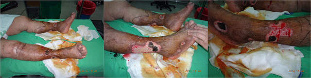

經過兩個月的抗凝血劑治療,及一般簡單換藥後,傷口已完全癒合。
Discussion:
靜脈性壞疽導因於靜脈拴塞,此疾病必須先排除調動脈性拴塞。導致原因可能為先天性凝血異常(常見如protein C deficiency, protein S deficiency, antithrombin III deficiency),或者外傷、感染、服用避孕藥、年老、或惡性腫瘤。因先天性問題多發生於年輕人,此患者靜脈性壞疽的原因極可能是感染及年紀大。治療的第一選擇為使用抗凝血劑,建議先使用先低分子量肝素,接著改為口服warfarin,外科血栓移除術只建議在發生深層靜脈拴塞合併腔室症候群時才使用。
附上Abstract原文如下:
Venous Skin Gangrene Patches Treated Successfully with Thrombolytic Therapy
Introduction
Venous gangrene is a rare conition. We report a case of venous gangrene, treated successfully with thrombolytic therapy.
Case report
A 77 years female with past history of hypertension, atrial fibrillation and peptic ulcer presented with pain and multiple skin dry gangrene over bilateral lower leg for the last 4days. There was no trauma history. She was sent to our emergence department due to fever and was admitted to medical service under the impression of cellulitis . Because the fever about 38-39C has persisted for 1week in vain with antibiotics treatment, surgeon was consult. When we examined the patient, she was able to walk, had multiple areas of dry gangrenous patches involving around 10% of nil lower legs and feet. All peripheral pulses were well palpable. The Doppler sonography showed no deep vein thrombosis.
WBC:10220, Seg:75, Lymph:18, Eosin:3,Hb:9.3, plate:391K, MCV:90,BUN/Cr:39.5/1.7,CRP:21.11
Patient received debridement 10 days after admission and only full thickness skin is involved. The fever subsided after operation. However, vomiting developed 2 days after operation. The panendoscopy showed gastric ulcer and duodenal ulcer. 4 days after operation, consciousness change, poor urine output and unstable hemodynamic state developed, so she was transferred to IC for intensive care. Brain CT showed dilatation of bilateral ophthalmic veins. CSF study showed gram positive coccus (oxacillin resistance staphylococcus aures was noted 3days later, the same with wound culture result). CNS infection was suspected. She was transferred to ward 6 days later. But after 5more days, she was transferred to ICU again due to CO2 retention with respiratory failure. Seizure attacked 10days later(POD25) when neurology doctor suspected cerebral vein sinus thrombosis(CVST) after review of the brain CT and history.
PT:12.9 sec (8-12),PTT:29.4 sec (24-36) Inr:1.22, RPR: 2X(+) ,TPPA:1:320X(+), Homocysteine:17.17umol/L (4.44-13.56), Protein C:37.8% (70-140), Protein S:17.5% (60-130), Antithrombin:60.9% (>75), Phospholipid Ab: 0.980RU/ml (<12(-)), Cardiolopin IgG:6.2 GPL (<15), Cardiolipin IgM :4.7MPL (<12.5),
Thrombophilia induced CVST and venous gangrene is suspected. The old age or infection may be the cause of thrombophilia.
Then antithrombotic therapy start. She was put on low molecular weight heparin (LMWH) for five days and also on oral anti-coagulants, monitoring INR, which was maintained between 2-3. The wounds start improving. The wound healed 2months later.
Discussion
Venous gangrene is a rare complication of venous thrombosis. It results from extensive venous occlusion in which the arterial tree remains patent. The diagnosis can be made with certainty only if the arteries are shown to be patent by examination of arteriography or Doppler sonography. Contrast venography(or MR venography) was the definitive test for the diagnosis. The most frequent manifestation of venous thrombosis is deep vein thrombosis, followed by phlegmasia alba dolens, phlegmasia cerulean dolens and venous gangrene.
Nonsuppurative thrombophilia may develop due to local trauma, prolonged inactivity, fungal infection, old age, pregnancy, the use of oral contraceptives, malignancy or caused by heparin-induced thrombocytopenia and thrombosis (HITT). Hereditary such as Protein C deficiency, protein S deficiency, Antithrombin III deficiency maybe considered if it develops in young adult. Suppurative thrombophilia may occur as a complication of iv line placement or iv drug abuse.
Anticoagulants should be given to try to control thrombosis and prevent pulmonary embolism-an important cause of death. Low Molecular Weight Heparin (LMWH) followed by oral wafarin is advised. Surgical thrombectomy to clear thrombus is recommended for patients with threatened limb loss or venous gangrene caused by massive DVT associated with high compartment pressures and arterial insufficiency (phlegmasia cerulea dolens).
沒有留言:
張貼留言